TRANSMISSION // 2611.23189v1
A shared memory for fixing medical image masks
Source published
GRACE retrieves verified examples to guide corrections to medical image segmentations. It helped most when input masks were weak, but transfer across modalities remains limited.
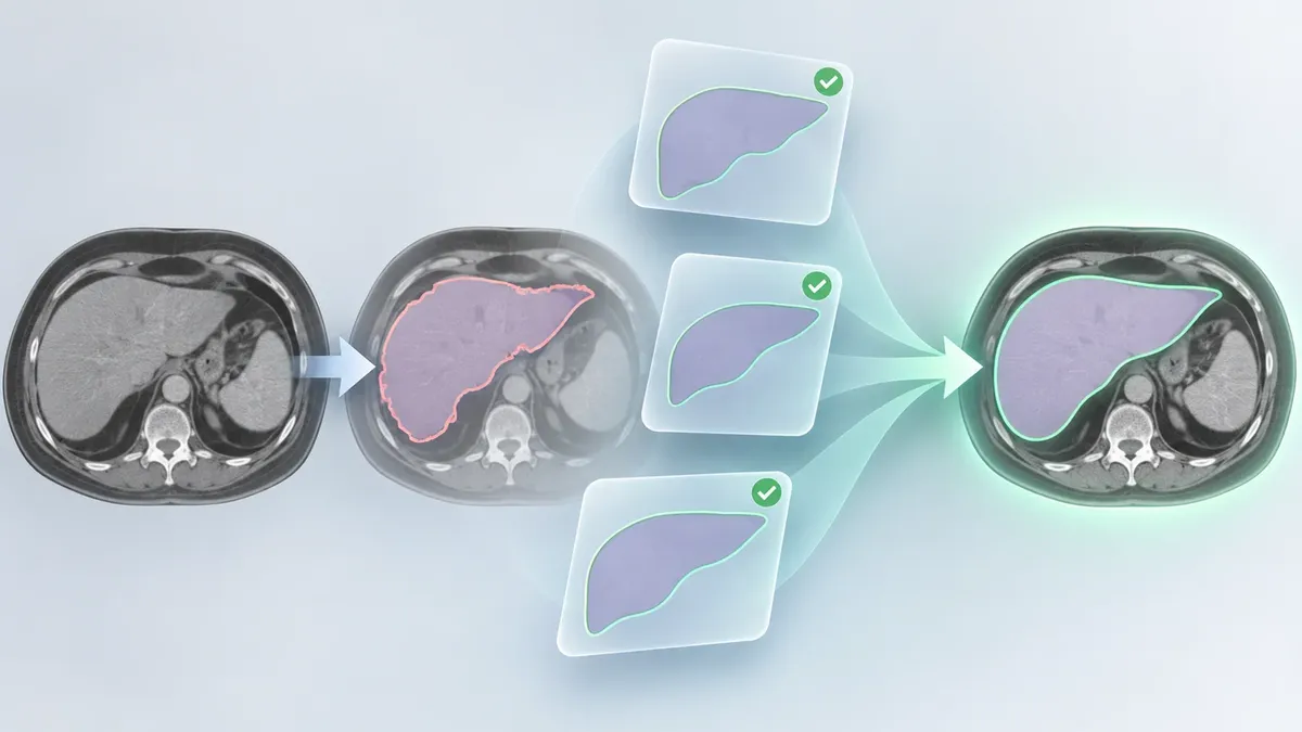
Medical scans are often divided into labeled regions—such as organs or lesions—before measurements can be made. But those computer-generated outlines can contain gaps, spill over boundaries or miss parts of a structure. A study of GRACE, a retrieval-guided correction system, asks whether one refiner can improve masks across many anatomies by consulting a shared collection of verified examples, rather than relying on a separately trained corrector for each target.
A segmentation mask is an outline drawn over an image to mark a structure of interest. In medical imaging, these masks can support quantitative analysis, but errors in their shape may need to be fixed before they are useful. Many correction systems are designed for one anatomy at a time. The authors of a study published in Transactions on Machine Learning Research propose another approach: let a general-purpose refiner consult a library of previously verified cases while editing a mask.
Their system, called GRACE, takes an image and an existing segmentation, retrieves examples judged similar, and uses one retrieved mask as a spatial guide for correction. Its knowledge base stores verified masks alongside image-and-mask representations. A frozen multimodal encoder creates the representations; a separate learned projection adjusts similarity rankings to better reflect agreement in mask shape. The refiner itself is trained once on data covering multiple organs and structures. The authors tested three versions, including a 3D U-Net that makes a direct correction and latent-space models that work through a compressed representation.
The evaluation covered 18 public datasets spanning CT, MRI and ultrasound. The training corpus included 15 datasets, and three others were held out for a dataset-transfer test. Seven upstream segmentation systems represented weak, intermediate and strong starting points. The team compared nine refinement configurations, including GRACE variants and other approaches. For transfer to the held-out datasets, those datasets’ cases were excluded from the knowledge base; the system used a fixed index built from training data. The researchers also ran a separate experiment in which verified examples of a previously unseen anatomy were added to the index without updating the refiner’s parameters.
The clearest gains appeared when the original masks were poor. For the weak tier, the input Dice similarity coefficient (DSC)—a measure of overlap between a predicted mask and a reference—was 0.386, and GRACE added as much as 0.35 DSC in the reported configuration. On a strong segmentor with an input DSC of 0.932, the average change was small. This pattern suggests that a retrieved shape example can be especially useful when the initial mask provides little reliable structure, while there is less room to improve an already accurate outline.
On the three held-out datasets, both GRACE variants improved DSC for each tested dataset-and-segmentor pairing. But this was not a test on entirely unfamiliar anatomy: those anatomies and imaging modalities were represented elsewhere in the training corpus. The authors describe it as transfer across datasets and acquisition conditions, not zero-shot generalisation.
The unseen-anatomy experiment offers a different, limited result. Adding verified support masks for a held-out organ improved performance without changing the refiner’s parameters. It required 118 to 302 support masks, however, and the authors caution that this does not demonstrate zero-shot ability. Their separate test of a completely held-out imaging modality was mixed: refinement transferred partially from CT to MRI, but failed on ultrasound in the reported setting. The ultrasound test also differed in image structure and view, so the result should not be read as a clean measure of modality alone.
There are other reasons for caution. GRACE’s benefit depends on having useful, representative examples in its knowledge base, and a wrong anatomy label can lead to a less helpful retrieved mask. Although average changes on strong inputs were small, some already-good masks became worse: among cases with input DSC above 0.8, 3.8% lost more than 0.05 DSC. The paper also reports that its slower, generative variant did not improve aggregate accuracy over the deterministic latent version, though it can provide sample-level uncertainty estimates.
These are benchmark results, not evidence that GRACE is ready to make clinical corrections without oversight. The study evaluates public datasets and reference masks; it does not establish improved patient outcomes or show how the system would perform in a live clinical workflow. The authors report publication in Transactions on Machine Learning Research in October 2026, so this is not merely an unreviewed arXiv submission. Still, results from the tested datasets do not settle how well retrieval-based correction will work across hospitals, rare conditions or other settings. The work’s central proposal is narrower: a shared, expandable memory of verified shapes may help one refiner handle diverse segmentation errors, especially when its starting mask is weak.
- medical image segmentation
- image refinement
- retrieval-augmented learning
- GRACE
- medical imaging AI
- Dice coefficient
Related reading
Automatically matched by shared topic keywords. These links do not imply that the sources agree or establish a consensus.