TRANSMISSION // 2611.14018v1
AI checks chest X-rays globally, then inspects regions to explain its reports
Source published
ARIVE first surveys a chest X-ray as a whole, then examines selected anatomical regions while forming a report. In tests using two MIMIC datasets, it scored well on measures of clinical content. A small expert review suggests its reasoning traces may help reviewers spot errors, but took longer. The work is an arXiv preprint, and the results do not establish performance in routine clinical practice.
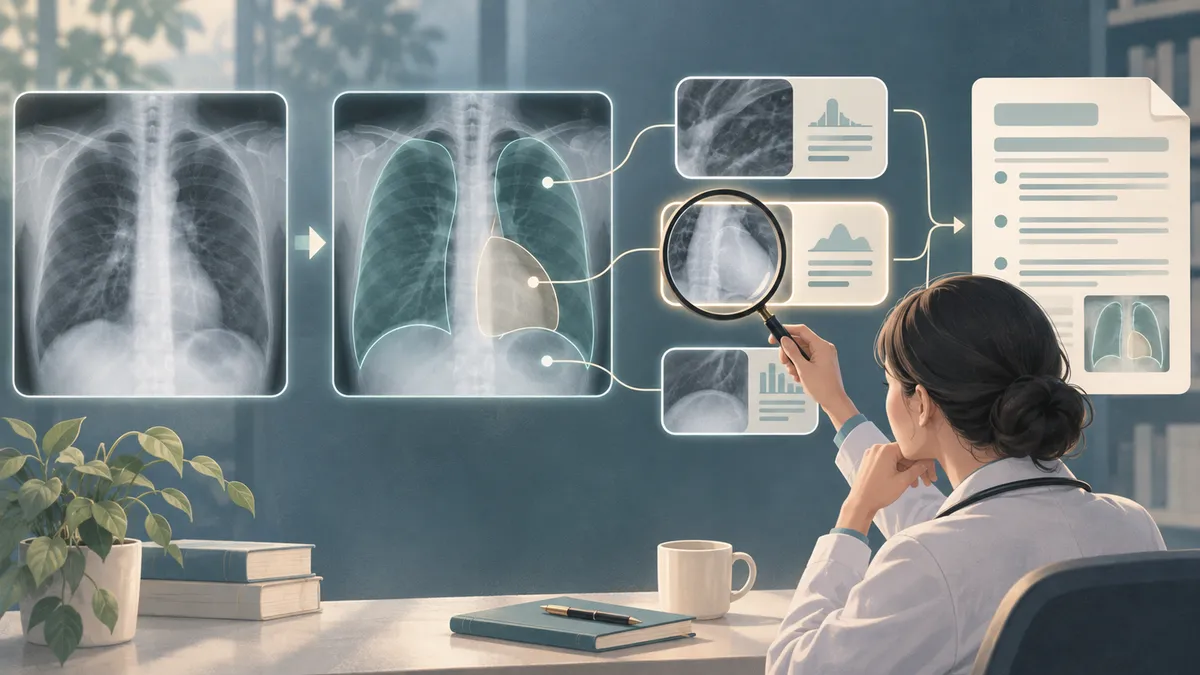
A fluent radiology report does not necessarily show which parts of an image support its claims. Researchers developed ARIVE, a system that first surveys a chest X-ray and then revisits selected anatomical regions while reasoning about what it sees. The aim is to make the path from image to report more inspectable—not to make AI interpretation self-validating.
Automated systems can produce plausible descriptions of chest X-rays, but a reader may not be able to tell what visual evidence led to a particular finding. Huang and colleagues propose a way to expose more of that path: have the model survey the whole image, inspect selected anatomical regions, interpret their visual evidence and then compose a report.
The framework, called ARIVE, is designed for chest radiographs. During generation, it first takes in the image as a whole, then requests selected anatomical regions. It encodes those local views and feeds them back into the model before it continues its interpretation. The resulting sequence records region requests, intermediate statements and the final report, so a reviewer can trace a report claim to earlier steps and associated image regions.
How the system was trained and tested
The researchers built 159,416 multimodal reasoning trajectories from the MIMIC-CXR dataset. They started with existing radiology reports and used a large language model to reorganize their findings into stepwise narratives. They then attached image-region boxes from Chest ImaGenome annotations, covering 29 predefined anatomical regions. These are report-derived training examples, not direct records of how a radiologist actually reasoned through each case.
ARIVE was first trained to produce the trajectories and reports. The team then applied an optimization method called Anatomical and Clinical-Content Optimization (ACCO), which rewards anatomical grounding and agreement with clinical content in both intermediate reasoning and the final report. Some rewards give extra weight to less common findings or regions.
The evaluation used MIMIC-CXR and MIMIC-ABN, an abnormality-focused subset of MIMIC-CXR. The authors compared ARIVE with six other systems using automated measures of clinical findings, relationships between findings and report errors. They also tested anatomical localization, asked an experienced radiologist to grade a sample of reports and reasoning traces, and compared review of reports with and without the traces.
What the study found
On both datasets, ARIVE scored higher than the compared models across the reported clinical-efficacy measures. On MIMIC-CXR, it led on all seven such measures; on MIMIC-ABN, it led on all six tabulated measures. The lead was not statistically significant in every comparison, and ARIVE did not lead on measures of wording overlap with reference reports. That distinction matters: matching clinical content and matching a reference’s phrasing are different goals.
The authors’ training comparisons suggest that regional image evidence and clinical-content optimization played complementary roles. Adding text-only reasoning did not consistently improve report quality. Interleaving regional inspection improved most clinical-efficacy measures compared with text-only reasoning, though one main CheXbert score dipped slightly on MIMIC-CXR. Adding ACCO to that setup improved all six tabulated clinical-efficacy measures on both datasets.
In blinded grading of 100 cases, ARIVE’s average report score was similar to Qwen3.5’s and higher than PriorRG’s, while the reference reports received the highest average. The radiologist rated ARIVE’s reasoning traces highly for diagnostic traceability. In a separate paired review, the radiologist identified errors in 11 reports when traces were available, compared with 8 without them. Median review time also increased, from 24.5 to 42.5 seconds per report. This is a preliminary result from one reviewer and a limited sample, not proof that traces improve clinical outcomes.
What remains uncertain
A visible reasoning trail is not the same as a correct one. The paper describes an unsupported finding that appeared in an intermediate step and carried through to the final report. The authors caution that generated reasoning should be checked against the image, rather than trusted because it is detailed or coherent.
The study also has important scope limits. Its evaluation used frontal chest X-rays from a single-institution dataset; it does not establish how the approach performs across hospitals, reporting styles, other imaging views or other body regions. The trajectories were reconstructed from reports and may inherit omissions, local conventions or rewriting errors. Anatomical supervision was sparse for some regions, and localization there remained weaker. Expert assessments involved small samples and a single radiologist.
The paper is an arXiv preprint, not evidence of established clinical effectiveness. Overall, ARIVE offers a way to make parts of AI-generated chest X-ray reports more inspectable. Whether that visibility leads to safer or more useful reporting in real clinical workflows remains to be tested.
- radiology AI
- chest X-rays
- report generation
- explainability
- medical imaging
- clinical evaluation
Reviewed cross-references
These connections were reviewed by a model, not independently fact-checked by a human. Similarity does not imply agreement; consult the original papers and the evidence below.
When a Chest X-ray Leads AI to Invent Findings in a Photograph
Supporting evidence: Both papers show that an inspectable or coherent diagnostic account need not be correct: ARIVE’s authors report that an unsupported finding can propagate through its reasoning trajectory into a report, while the Trick-or-Treat study found that mismatched radiographs could steer three models to hallucinate matching findings in synthetic clinical photographs. These results support the bounded caution that reasoning traces or multimodal context do not guarantee evidential grounding; the tasks, image inputs and evaluations differ, and Trick-or-Treat does not evaluate ARIVE.
Model review: · openai-codex/gpt-6-luna
Evidence and provenance
Selected excerpts, not complete papers. PDF extraction can omit figures or damage equations, tables and reading order; sources may be preprints.
2611.14018v1 — Original paper
Excerpt reference:
static/papers/2611.14018v1.md:1259-1305
Basis: Paper text
Source SHA-256:59f05fde63bb9bea6e7afc61f2c1728b848ba5d4cfc5d59e9660b51c444d61bd2611.06631v1 — Original paper
Excerpt reference:
static/papers/2611.06631v1.md:417-476
Basis: Paper text
Source SHA-256:627a5484eb79f83cd2e43e94b310b2fc62331e48318dae1588605e343925d5662611.06631v1 — Original paper
Excerpt reference:
static/papers/2611.06631v1.md:477-510
Basis: Paper text
Source SHA-256:627a5484eb79f83cd2e43e94b310b2fc62331e48318dae1588605e343925d566
Related reading
Automatically matched by shared topic keywords. These links do not imply that the sources agree or establish a consensus.
Why more AI tools alone may not deliver complete radiology reports
Source published
Shared keywords: medical imaging
Can one medical-image detector learn across X-rays and mammograms?
Source published
Shared keywords: chest x rays, medical imaging
Mammography AI heat maps perform worse in dense breasts—but the reasons matter
Source published
Shared keywords: medical imaging
VoluCompose compresses medical scans for AI while preserving benchmark performance
Source published
Shared keywords: medical imaging
An AI report can copy the structure and still get the facts wrong
Source published
Shared keywords: report generation