TRANSMISSION // 2611.10739v1
Pulse shape changes before stroke recognition—but an early-warning system remains unproven
Source published
A retrospective preprint finds stroke-related patterns in hospital pulse recordings across two datasets. Strong headline classification scores come with important limits: recognition is not stroke onset, patient-grouped discrimination was weak, and short recordings sharply reduced alert coverage.
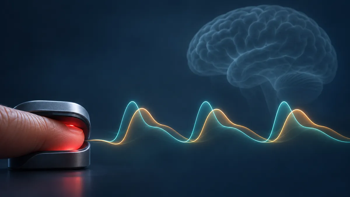
A bedside pulse sensor records more than heart rate and oxygen saturation. The shape of each pulse also reflects changing circulation. Could those usually overlooked contours help identify patients who need a neurological examination?
Looking beyond the pulse count
A new preprint by Cheng Ding and colleagues investigates whether the peripheral pulse carries a detectable signature before clinicians recognise a stroke. The study finds reproducible patterns in hospital recordings, but does not establish that a device can predict a stroke before it begins.
The researchers analysed photoplethysmography, or PPG: an optical measurement of blood-volume changes recorded by pulse oximeters and many wearables. Each beat produces a rising and falling contour. Its timing, amplitude and variability reflect several interacting influences, including cardiac contraction, arterial properties and vascular tone.
The central question was whether those contours differ between earlier recordings and the period approaching documented stroke recognition in the same patient.
Two hospitals, one important timing distinction
The warning analysis included 176 patients from the MIMIC-III intensive-care resource and 154 from Stanford's MC-MED emergency-department resource. Both datasets were retrospective. Candidate event times were extracted from clinical notes with language-model assistance and adjudicated by physicians.
These timestamps mark documented recognition, not necessarily the biological beginning of a stroke. Sedation, confusion or subtle symptoms can delay recognition. Consequently, a waveform recorded before the timestamp may already reflect a stroke underway, its systemic consequences or treatment-related changes.
The researchers extracted 17 descriptors of pulse shape and supplied ordered sequences to a one-dimensional residual neural network. Models developed in MIMIC-III were applied to MC-MED without retraining. The populations differed substantially: every intensive-care warning patient had an admission-level haemorrhagic diagnostic code, while the emergency cohort was predominantly ischaemic.
What the headline scores measure
At the nominal 360-minute setting, the original analysis reported F1 scores of 0.941 in MIMIC-III and 0.989 in MC-MED, with sensitivities of 0.998 and 0.980. F1 combines precision—how many positive classifications are correct—with sensitivity—how many labelled positive windows are detected.
Those numbers describe classification of recording windows, not the proportion of future strokes successfully predicted. The six-hour setting defined a broad pre-recognition interval; it did not require an alert exactly six hours before an event. Earlier windows provided the stable comparison, with buffers excluding observations around the class boundary and immediately before recognition.
The labels were also strongly imbalanced: warning windows predominated, increasingly at longer horizons. That makes high F1 insufficient evidence of useful discrimination. The original development grouped recording identifiers rather than ensuring that all recordings from each patient stayed together.
A stricter patient-grouped analysis produced a morphology F1 of 0.742 on 15,117 windows from 164 MIMIC-III patients. The authors also describe pooled patient-grouped discrimination as low. They suggest that personal changes may matter more than differences between people, but explicitly acknowledge that a weak signal could explain the result too; within-patient discrimination was not directly measured.
Which parts of the pulse mattered?
Feature-attribution analysis highlighted crest time: the interval from the start of a pulse to its systolic peak. Its deviation from a patient's baseline was especially influential. Feature-importance rankings agreed across the two cohorts, with a Spearman correlation of 0.865.
On identical patient-grouped windows, morphology exceeded heart rate alone by 0.123 in F1. Against heart rate plus heart-rate variability, the advantage narrowed to 0.049, with a confidence interval including zero.
Attribution identifies what influenced a model, not what caused a stroke. A small analysis combining ECG and PPG suggested that the relationship between crest time and heart rate might weaken before recognition. Its uncertainty was wide, and the researchers treat that mechanism as a hypothesis rather than an established finding.
Turning scores into alerts
False positives were common in high-risk patients without documented stroke. At the six-hour setting, approximately 29% of control windows in MIMIC-III and 14% in MC-MED were positive. Requiring five consecutive positive windows reduced the proportion of control patients or patient groups receiving an alert to about 17% and 2%, respectively.
A separate available-recording analysis shows why window sensitivity is not patient coverage. The persistence rule alerted before recognition in 168 of 174 intensive-care patients, but only 46 of 112 emergency patients. Sixty-two emergency patients had fewer than five windows, making confirmation impossible.
Among alerted patients, median intervals to recognition were 5.25 and 1.68 hours. These are retrospective intervals conditional on an alert, not demonstrated prospective warning times.
A candidate signal, not a clinical alarm
A separate model using the first two hours of PPG after admission or emergency arrival classified worsening or death versus improvement with F1 scores of 0.714 and 0.691. That concerns subsequent clinical course, not impending stroke.
Unmodelled influences—including peripheral perfusion, motion, probe placement and treatment—could affect the pulse patterns. Both resources came from US health systems, and wearable use was not tested. The findings support further investigation of pulse morphology, but prospective evaluation must establish whether alerts identify actionable neurological changes without an unacceptable alarm burden.
- photoplethysmography
- stroke recognition
- pulse waveform morphology
- clinical artificial intelligence
- patient-grouped evaluation
- false alerts