TRANSMISSION // 2611.09058v1
Prostate ultrasound AI uses past cases to explain predictions—and flags disagreement
Source published
ProtoCouncil compares prostate micro-ultrasound images with anatomical regions from real training cases. In a preprint, researchers report improved cancer-detection scores across three cohorts, while disagreement between its three models identifies less reliable predictions. Benefits for clinical decisions remain untested.
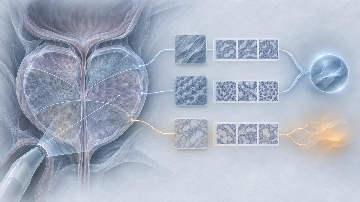
An AI prediction is more useful if clinicians can inspect its supporting evidence and recognize when it may be wrong. A new prostate imaging study explores both goals by combining three models that compare ultrasound images with previously examined cases.
ProtoCouncil, described in a November 6 preprint by Mohammad Mahdi Abootorabi and colleagues, combines prostate cancer detection with a traceable scoring system. Instead of producing only a cancer probability, it connects its scores to anatomical regions from real training cases. When its three component models disagree, it can withhold the prediction. The paper is under review; its findings do not establish clinical benefit.
Learning from imperfect biopsy labels
Micro-ultrasound produces real-time prostate images, but recognizing suspicious tissue depends heavily on clinical experience. Training AI to interpret these images presents another difficulty: biopsy pathology describes a sampled tissue core, not the exact location of cancer in the corresponding ultrasound image.
The researchers used 8,606 biopsy cores from 846 patients across three cohorts. Each core had an ultrasound frame acquired before needle firing, alongside masks identifying the prostate and needle trace. Models also received clinical information, including age, prostate-specific antigen measurements and core location.
The development cohort contained 6,588 cores from 693 patients. The team used five-fold cross-validation with patients kept separate between training and validation. Two external cohorts, containing 111 and 42 patients, tested transfer without retraining on those cohorts.
The study defined clinically significant prostate cancer as Grade Group 3 or higher. Its reported results should therefore be understood using that specific threshold.
Three models, anatomically anchored examples
ProtoCouncil uses MedSAM, DINOv3 and V-JEPA 2.1, pretrained respectively for medical-image segmentation, natural-image representation learning, and image-and-video representation learning. Each was adapted to prostate cancer detection before its features were used to train a prototype-based classifier.
These prototypes represent three anatomical families: the whole gland, the needle track within the gland, and six gland sectors. Each encoder has 36 prototypes spanning benign tissue, Grade Groups 1–2 and Grade Groups 3 or higher.
After training, the prototypes are replaced with representations of actual training regions from distinct patients. For cancer prototypes, candidate cores must have at least 40% tumor involvement. This gives each prototype an identifiable source case, although that core-level pathology does not establish exactly where cancer lies within every represented region.
Each diagnostic class score is a baseline value plus weighted similarities to its prototypes. Combining the three encoders preserves this additive structure, allowing contributions to be traced through the final score. The combination weights are estimated without pathology labels, but the underlying cancer detectors and prototype classifiers are trained with labeled data.
Stronger ranking, with important qualifications
For clinically significant cancer, ProtoCouncil achieved an area under the receiver operating characteristic curve, or AUROC, of 82.9% in development cross-validation. AUROC measures how well scores distinguish positive from negative cases across thresholds; it is not the percentage of diagnoses that are correct.
The result exceeded the strongest previously published comparator, Prost-RL, by 4.3 percentage points. The reported 95% confidence interval for that difference was 1.4–7.3 points.
External clinically significant cancer AUROCs were 80.0% and 79.5%, the highest point estimates among the evaluated methods. However, advantages over a separately fine-tuned DINOv3 comparator were smaller: 0.5 and 1.8 points, respectively.
Performance also depended on the task. All-cancer AUROC fell to 63.6% in the smaller external cohort, where most cancer cores were Grade Groups 1–2. Patient-level results did not consistently lead the comparators, and differences from the strongest methods were not statistically significant. Clinicians’ recorded PRI-MUS risk scores also retained a higher clinically significant cancer AUROC in the available external comparison.
Disagreement identifies a harder subset
For selective prediction, the system retained a call only when all three encoders agreed. Decision thresholds were established on development validation data at 80% specificity and then applied unchanged externally.
Across the external cohorts, fused predictions were correct in 78.6% of cores when no encoder opposed them. Accuracy dropped to 27.0% when two encoders opposed the fused call.
Withholding disagreements retained 58% and 54% of cores in the two external cohorts. Balanced accuracy—the average of sensitivity and specificity—rose by approximately 7 and 15 percentage points among those retained. This improves performance on a selected subset, not on every core: almost half still require another assessment pathway.
Traceability is not yet clinical validation
The nearest same-class gland examples had more similar tumor involvement than average same-class examples, suggesting that prototype similarity captures some relevant tissue information. That exploratory finding does not prove that the explanations help clinicians.
All images came from the same micro-ultrasound platform, and the smallest external cohort included only six patients with clinically significant cancer. One author disclosed an executive role at the device manufacturer. Whether displaying supporting and conflicting cases improves clinical decisions remains an explicit question for future studies.
- prostate cancer
- micro-ultrasound
- ProtoCouncil
- interpretable AI
- prototype learning
- foundation models
- selective prediction
Related reading
Automatically matched by shared topic keywords. These links do not imply that the sources agree or establish a consensus.
A Low Error Rate Can Still Hide a Huge Ultrasound Measurement Mistake
Source published
Shared keywords: selective prediction
Reading the warning signs: an explainable way to stop failing web agents early
Source published
Shared keywords: interpretable ai
When a smaller AI model cannot make a query cheaper
Source published
Shared keywords: selective prediction
Learning Payment Fraud Sequences to Train Better Detectors
Source published
Shared keywords: interpretable ai
AI moral choices shift when models take a role in a video game
Source published
Shared keywords: foundation models