TRANSMISSION // 2611.08969v1
PhysioWave-T compresses biosignal inputs without expanding the token count
Source published
A preprint describes wavelet-based AI models that shorten the sequences used to analyze brain, heart and muscle recordings. Benchmark results suggest substantial memory savings, but seizure-monitoring gains depend on the evaluation metric and remain short of clinical validation.
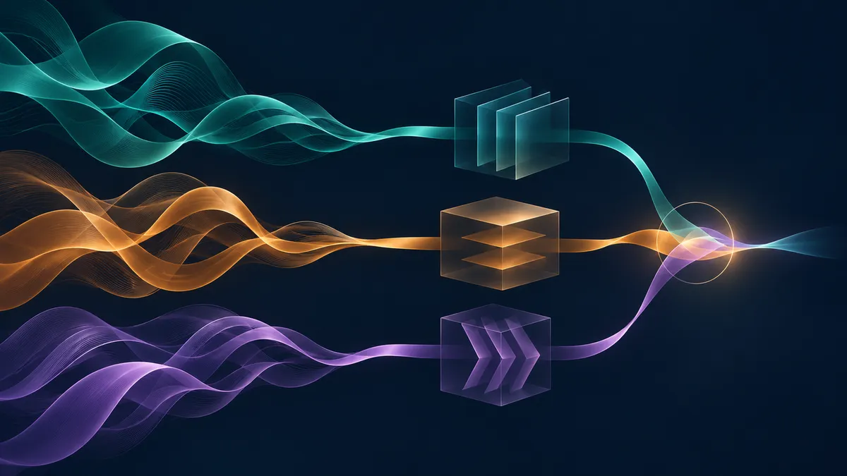
Can AI examine physiological signals at several timescales without turning every frequency band into extra computational work? PhysioWave-T combines those bands before a Transformer processes them, while adapting its input layers to different recording setups.
Brain, heart and muscle recordings contain patterns at different timescales, but capturing that detail can make AI models expensive. More electrodes, longer recordings and additional frequency bands all increase the amount of information to process. A preprint by Yanlong Chen and colleagues proposes PhysioWave-T, a family of models designed to preserve useful multiscale information while shortening the sequences passed to a Transformer.
The manuscript, submitted to arXiv on November 6, 2026, reports experiments on electroencephalography, or EEG; electrocardiography, or ECG; and surface electromyography, or sEMG. These measure electrical activity associated with the brain, heart and muscles, respectively. The results are research benchmarks, not evidence of a clinically validated monitoring system.
Combining scales before making tokens
The central technique builds on wavelets: filters that separate a changing signal into components associated with different timescales. In the authors’ earlier approach, each component became a separate sequence of tokens, the numerical units processed by a Transformer. With three decomposition levels producing four components, that meant four times as many tokens.
PhysioWave-T instead uses “dynamic scale folding.” It combines the components into one signal per channel before creating tokens. A small learned network assigns weights using local signal statistics, while learned synthesis filters refine the combination. The model can therefore change which scales it emphasizes across channels and time intervals.
This is learned compression, not a guarantee that every detail survives. Its practical justification comes from the experiments: on two EEG tasks, dynamic folding slightly outperformed the longer, scale-expanded representation. Simply averaging the components performed worse, indicating that the learned combination mattered.
Across four EEG configurations, the authors report Transformer latency reductions of 4.0–7.2 times and peak-memory reductions of 6.8–12.7 times compared with scale expansion. Profiling used an NVIDIA A100 with matched batch sizes. These are comparisons against a particular alternative representation, not speedups over every competing model or measurements on wearable hardware.
Different sensors, shared backbones
Recording setups vary substantially. PhysioWave-T handles this through configuration-specific input layers, selected using channel count and sampling rate, followed by a shared Transformer within each signal type. Learned channel-name embeddings identify which electrode or lead produced each input.
There are separate EEG, ECG and sEMG encoders—not one universal model trained jointly across all three. An unfamiliar configuration can require new input layers and channel embeddings trained for the downstream task; compatibility does not mean immediate use without adaptation.
The EEG encoder was pre-trained on approximately 15.4 million four-second windows spanning 19–128 channels. Training hid 70% of selected patches in the raw input, then asked the model to reconstruct both the measured signal and its folded-wavelet representation. This self-supervised task learns from recordings without requiring a diagnostic label for every window.
After fine-tuning, the EEG model led eight of nine reported metrics against four selected baselines across sleep staging and two tasks involving responses to events. Benefits from pre-training varied: the sleep-staging architecture was already strong when trained from scratch, whereas detecting error-related responses benefited more from adapting pre-trained weights.
The authors also report leading gesture-recognition results among the compared sEMG models and the highest macro-F1 on two ECG benchmarks. Performance was not uniformly best: other models retained higher ECG ranking scores on some datasets.
Better fusion depends on the question
A second component, reliability-aware latent fusion, or RALF, combines compact summaries from the independently trained encoders. It estimates each modality’s reliability for each window and reduces the contribution of degraded inputs. Training includes synthetic noise, drift, saturation, bad channels and dropout, alongside missing-modality examples.
On SeizeIT2, the reported test set contained 29 patients, 2,912 recording hours and 181 seizures. RALF increased window-level area under the precision–recall curve from 0.321 for the best single-modality model to 0.399. This metric summarizes the tradeoff between finding positive windows and avoiding incorrect positive predictions.
Event detection tells a more qualified story. At 11 false alarms per hour, the detailed results report 74.4% seizure sensitivity for RALF, versus 61.4% for EEG alone—but 78.9% for simpler weighted-logit fusion. The introduction inconsistently attributes 78.9% to RALF; the abstract and detailed results give 74.4%. RALF was therefore not best at that operating point.
Eleven false alarms per hour remains a substantial burden. The authors also acknowledge that their corruption tests are synthetic and identify prospective recordings with natural sensor failures as future work. Their own experiments report three-seed means, while some comparator results come from earlier publications. Finally, the supplied PDF extraction contains visibly garbled figure material; this account relies on readable text, tables and captions, not independently inspected plots. The evidence supports a promising efficiency strategy, not deployment readiness.
- PhysioWave-T
- biosignals
- wavelets
- Transformers
- EEG
- ECG
- surface electromyography
- multimodal fusion
- seizure detection