TRANSMISSION // 2611.08771v1
Federated AI generates brain MRI scans with a diversity trade-off
Source published
A preprint comparing federated and centralized training found similar diagnosis-related performance in synthetic brain MRI. Federated samples scored closer to real scans on one metric but were less diverse, and neither approach reproduced the real-data distribution.
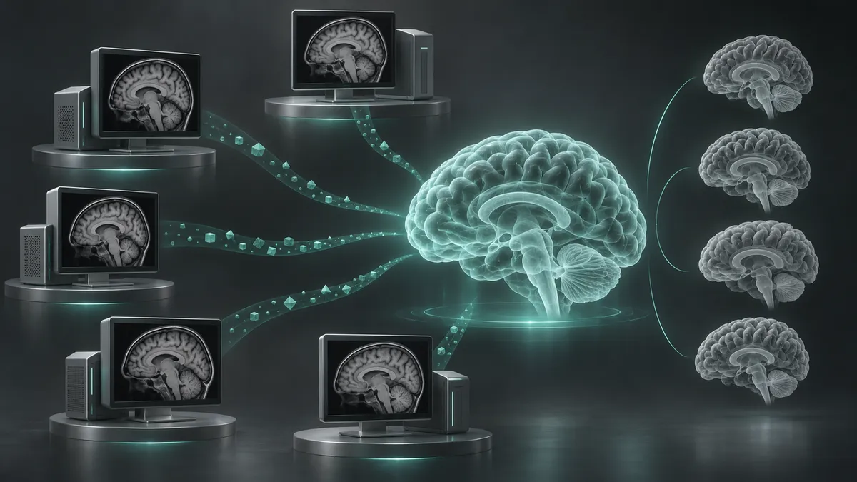
Can researchers train a brain-image generator without collecting everyone’s scans in one place? A controlled comparison suggests they can achieve results comparable to pooled-data training—but with an important qualification: the federated model produced a narrower range of synthetic brains.
Comparing two ways to train
Abhijith Shaji and colleagues compared two training arrangements for the same three-dimensional brain MRI generator. Centralized training brought the scans together. Federated training kept scans at five separate data sites and combined locally trained model parameters instead. Their report, posted to arXiv on November 5, 2026, is a preprint; the supplied record does not establish peer review.
The motivation is practical. Brain-imaging datasets can be small and unevenly distributed between diagnoses, while consent and governance restrictions complicate pooling. Synthetic scans might help supplement such datasets. This study tested image generation, however, not whether adding generated scans improves a clinical system.
What data and models were used?
The researchers used T1-weighted MRI from the Alzheimer’s Disease Neuroimaging Initiative and the National Alzheimer’s Coordinating Center. Three ADNI study phases and two NACC scanner groups formed the five federated sites. These were dataset groupings, rather than simply five individual hospitals.
There were 7,321 diagnosis-labeled training scans, classified as cognitively normal or Alzheimer’s disease. Training, validation and test participants were separate, preventing repeated scans of the same person from crossing those boundaries. ADNI participants could contribute multiple longitudinal scans. Images were processed into a common orientation, template and resolution.
Both pipelines used two stages. First, an autoencoder learned to compress MRI volumes into smaller numerical representations and reconstruct them. Next, a latent diffusion model learned to generate those representations by removing added noise, with the requested diagnosis guiding generation. The autoencoder then converted the result into an image.
The comparison used matching architectures, losses and sampling procedures. During federated training, every site trained locally for one epoch per round, and the server averaged parameters according to each site’s scan count. Scans, compressed image representations and labels stayed local. Shared information included model parameters, autoencoder optimizer statistics and summary numbers needed to calculate a common scaling factor.
How the images were evaluated
Each model generated 1,500 cognitively normal and 1,500 Alzheimer’s-labeled volumes. Researchers compared these with 1,407 real validation and test scans, using paired random seeds for generation.
Three measures addressed different questions. A classifier trained on real scans assessed whether synthetic images expressed their intended diagnosis. Multiscale structural similarity, or MS-SSIM, measured similarity between images within each group: higher scores meant less diversity. Fréchet Inception Distance, or FID, compared distributions of image features extracted by a pretrained medical-imaging network. Lower FID meant greater similarity to the real reference in that feature space, not necessarily greater clinical validity.
Comparable diagnosis signals, narrower variation
The autoencoders reconstructed real images similarly. Pooled validation reconstruction scores were 26.09 decibels for federated training and 26.30 for centralized training on the peak signal-to-noise ratio measure.
The diagnosis classifier achieved an area under the receiver operating characteristic curve, or AUC, of 0.934 on federated synthetic scans and 0.922 on centralized synthetic scans, compared with 0.920 on real held-out scans. These results concern recognition of labels in generated images. They do not show that synthetic data improved diagnosis in real patients.
Federated generation also had lower pooled FID: 1.143 × 10⁻³, versus 1.843 × 10⁻³ for centralized generation. Yet a sample of real training scans scored 0.0156 × 10⁻³ against the same reference—substantially closer than either synthetic set.
Diversity favored centralized training. For cognitively normal images, MS-SSIM was 0.918 for federated generation, 0.905 for centralized generation and 0.896 for real scans. Alzheimer’s images followed the same pattern. Federated samples were more alike, despite their better FID.
What the comparison leaves unresolved
The authors suggest that averaging updates across different sites may favor anatomy common to those sites, with the autoencoder potentially contributing too. These are proposed explanations, not experimentally isolated causes. Better FID therefore should not be read as an across-the-board improvement: a narrower distribution can score closer to typical real images while representing atypical brains less well.
The evaluation covers two diagnosis categories and these particular dataset groupings. It does not establish performance for other diseases, clinical usefulness or resistance to privacy attacks. Keeping scans local avoids transferring them, but the reported experiment does not demonstrate that shared updates or generated images cannot disclose information.
The supported conclusion is specific: federated training produced competitive synthetic MRI under the chosen measurements, without pooling scans, but sacrificed some diversity and remained measurably different from real data.
- federated learning
- brain MRI
- latent diffusion
- synthetic medical imaging
- Alzheimer’s disease
- data diversity
Related reading
Automatically matched by shared topic keywords. These links do not imply that the sources agree or establish a consensus.
In federated AI, participant selection matters under a strict training limit
Source published
Shared keywords: data diversity, federated learning
FedFiOT matches neurons to merge federated models with fewer exchanges
Source published
Shared keywords: federated learning
Small AI updates can still reveal private images
Source published
Shared keywords: federated learning
Can Communities Help Shape AI Without Handing Over Their Data?
Source published
Shared keywords: federated learning