TRANSMISSION // 2601.16759v2
A public image collection aims to give ERCP AI a clearer test
Source published
Researchers have released a curated collection of fluoroscopic images from endoscopic retrograde cholangiopancreatography (ERCP), a procedure used to diagnose and treat bile-duct and pancreatic conditions. The dataset includes images from 1,602 patients, with 5,519 labeled images reviewed by clinicians. A trial classification experiment shows that image-based prediction is feasible on this collection, but it does not establish clinical readiness or performance at other hospitals.
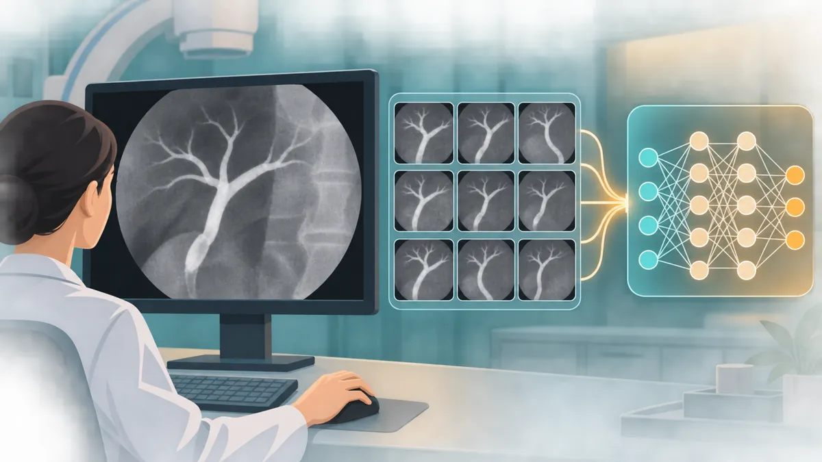
Medical AI needs more than clever algorithms: it needs suitable data on which different systems can be trained and fairly tested. For ERCP, a procedure guided in part by X-ray images, public image collections have been scarce. This paper introduces a dataset intended to help researchers study whether computers can classify relevant findings in those images.
Endoscopic retrograde cholangiopancreatography, or ERCP, is a procedure used to investigate and treat problems involving the bile ducts and pancreas. Clinicians use fluoroscopy—live X-ray imaging—to guide instruments and view contrast-filled ducts. Could machine-learning systems help analyze these images? One obstacle is a lack of publicly available ERCP image data that researchers can use to develop and compare methods.
A team led by Alda João Andrade and colleagues presents a curated collection intended to address that gap. The work is a data-resource paper: its central contribution is the dataset and the process used to prepare and label it, rather than a new diagnostic AI system.
From hospital archive to research dataset
The researchers retrospectively collected DICOM images from ERCP procedures performed at a tertiary-care hospital between 2015 and 2025. They converted the images to PNG and organized them into a raw collection and a processed collection. The raw set contains 19,018 images from 1,602 patients. Some source images showed several frames arranged together, so the team used image-processing methods to split these composites into individual sections. The resulting processed set contains 19,317 images; it is larger because some raw images yielded multiple sections.
The images came from different fluoroscopy systems and stages of procedures, including images used for orientation, contrast studies and therapeutic maneuvers. That variation may make the collection more representative of the kinds of images encountered in routine practice, but it also means that image appearance can vary for reasons unrelated to disease.
A subset of 5,519 images was labeled in categories including lithiasis (stones), bile leaks, benign and malignant strictures, and normal findings. The paper says two gastroenterologists performed the annotations, with review by a senior clinician and cross-checking against procedure reports. Images could also be marked to keep or discard, depending on whether they showed a useful diagnostic view. The remaining images are not labeled for these categories, so they should not be treated as confirmed examples of any particular finding.
The team describes anonymization procedures, including cropping and masking some image types to remove identifying borders, and replacing patient identifiers with numeric codes. The dataset and processing code are reported as publicly available through Figshare and GitHub.
A first test of classification
To demonstrate how the collection might be used, the researchers fine-tuned several image-classification models to distinguish the labeled categories. They divided images by ERCP exam so that frames from a patient would not be spread across training and test sets—a useful step to reduce the risk that a model appears successful by seeing near-related images during training and evaluation.
EfficientNet-B7 had the strongest reported result among the tested architectures: a macro F1-score of 0.738 on the test set. F1 combines precision and recall, and the macro version averages performance across classes rather than allowing more common classes to dominate the score. The result suggests that the images contain patterns a model can use for this classification task under the study’s test setup. It is not evidence that the model can diagnose patients reliably in clinical practice.
What the result cannot tell us
The experiment is a technical demonstration on data from one hospital, not an external validation across institutions, imaging systems or patient populations. Performance could change when images come from a different site or workflow. The paper’s reported scores also do not, by themselves, show how a model would affect clinical decisions, patient outcomes or procedure safety. No comparison with clinicians’ performance is described in the supplied evidence.
The labels are expert-reviewed, but they are still a reference constructed from image review and procedure reports, not an infallible measure of ground truth. The paper does not establish that every image in the collection is suitable for every possible research task. Researchers will need to inspect the dataset documentation, label distribution and patient-level splits for their intended use. The collection may support supervised learning with labeled examples and other approaches that also use unlabeled images, but those possibilities are not demonstrations of improved clinical care.
The paper is available as an arXiv preprint, a form of public research posting that may not have undergone peer review. The source text and retrieval material available here provide the paper’s account, but do not independently verify the dataset’s contents or download access. As with any medical-image resource, researchers should also consult its repository terms, privacy safeguards and documentation before use.
For now, the most defensible takeaway is modest: this is a potentially useful, openly described ERCP imaging resource, accompanied by an initial classification experiment. It gives researchers material with which to test ideas, while leaving the larger questions—generalization, clinical usefulness and safe deployment—to future studies.
- ERCP
- medical imaging
- artificial intelligence
- fluoroscopy
- clinical datasets
Related reading
Automatically matched by shared topic keywords. These links do not imply that the sources agree or establish a consensus.
Teaching medical AI to stop inventing findings can also expose what it misses
Source published
Shared keywords: medical imaging
Using generated ultrasound images to choose real ones worth labeling
Source published
Shared keywords: medical imaging
Mammography AI heat maps perform worse in dense breasts—but the reasons matter
Source published
Shared keywords: medical imaging
Can one medical-image detector learn across X-rays and mammograms?
Source published
Shared keywords: medical imaging
EvoML checks machine-learning workflows in stages and saves successful fixes
Source published
Shared keywords: medical imaging