TRANSMISSION // 2026.11.19.767918v1
How root cell structure relates to CYCB1 signals
Source published
A bioRxiv preprint links differences in Arabidopsis root-cell nuclear remodeling with CYCB1;1 reporter signals and G2 duration, cautioning against treating signal intensity as a universal measure of cell-cycle activity.
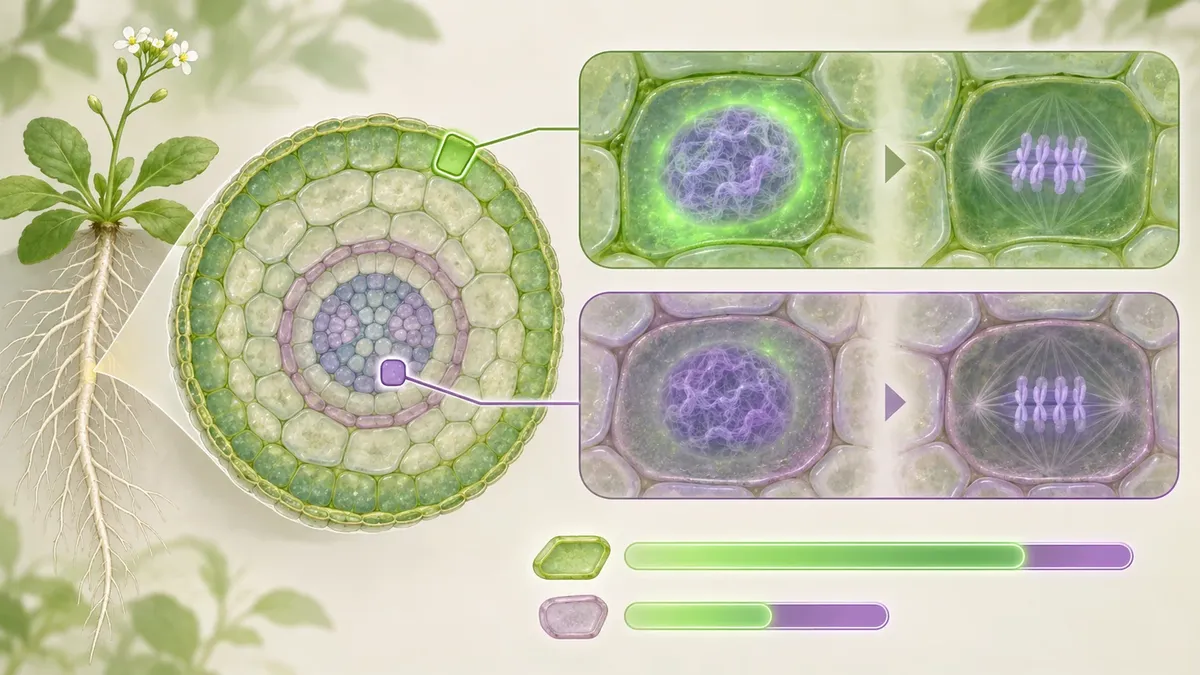
As plant cells prepare to divide, their chromatin—the DNA and its packaging—changes organization. A study of Arabidopsis roots compares these structural changes across tissues and examines how they relate to a commonly used cell-cycle reporter. The reported associations suggest that cell type matters when interpreting reporter signals, but do not establish what causes the differences.
A bright cell-cycle reporter signal is often used to track activity in plant tissues. But does the same signal mean the same thing in every cell type? In a bioRxiv preprint, Pasternak and Lebedev examine how nuclear and chromatin architecture across Arabidopsis thaliana root tissues relates to accumulation of the CYCB1;1 reporter. Their findings caution against treating reporter intensity as a universal quantitative measure of cell-cycle activity.
A structural transition
Before mitosis, a cell reorganizes its genetic material. During the transition from G2, a stage before mitosis, chromatin condenses into chromosomes and the nucleus changes substantially. The authors ask whether differences in a cell’s starting nuclear and chromatin organization are associated with this remodeling, CYCB1;1 reporter signals, and the timing of the G2-to-mitosis transition.
CYCB1;1 reporters are used to visualize cell-cycle activity in plants. The preprint emphasizes that their signal should not automatically be read as a direct count of dividing cells. It proposes that cell-type-specific architecture may be relevant to interpreting reporter accumulation, while noting that the mechanism remains to be established.
Comparing root tissues
The researchers studied Arabidopsis roots, comparing outer tissues—the epidermis and cortex—with inner tissues, including the endodermis and pericycle. They used three-dimensional imaging and image analysis to characterize nuclear and chromatin features. To visualize euchromatin, a relatively less compact form of chromatin, they used the histone marker H3K4me1. They also examined two reporter lines: CYCB1;1::GFP and CYCB1;1::GUS.
For the GFP reporter, seedlings were fixed and stained, and GFP signal was detected after immunolocalization with an anti-GFP antibody. Three-dimensional image stacks were used to quantify reporter signals; the methods report scanning at least 20 roots per variant. In one part of the chromatin analysis, cells undergoing mitosis were identified by a visible cell plate, helping the researchers compare cells at that stage.
What the preprint reports
Epidermal and cortical cells showed greater structural differences between G2 and mitosis than endodermal and pericycle cells. The outer tissues also had higher CYCB1 reporter signals, while the inner tissues had lower signals. In cells identified as mitotic, measured euchromatin geometry was broadly similar across the examined cell files, despite differences in chromatin organization between cell types.
The authors report that cells with greater structural separation between G2 and mitotic chromatin configurations also had higher CYCB1 reporter signals and longer G2 duration. Conversely, cells with smaller structural differences showed lower reporter signals and shorter G2 duration. This describes an association across cell types—not proof that greater remodeling causes a longer G2 phase or higher reporter accumulation. The authors say direct measurements of remodeling dynamics and cyclin-dependent kinase activity are needed to establish the underlying mechanism.
Interpreting the signal
The practical takeaway is a caution, not a rejection of the reporter. CYCB1;1 intensity should not be treated as a universal quantitative measure of cell-cycle activity across structurally distinct cell types. The study suggests that nuclear and chromatin architecture provides useful context for interpreting reporter measurements in intact tissues. It does not directly compare reporter brightness with division frequency, so it cannot show how well intensity predicts that rate.
There are limits to what can be concluded. This is a bioRxiv preprint and has not been certified by peer review. The work examines Arabidopsis roots under the conditions described in the paper; it does not establish that the reported relationships apply to other plants, organs, or environments. The proposed link between starting architecture and the physical requirements of mitotic entry remains a model, not a demonstrated causal mechanism. Further measurements will be needed to clarify how nuclear structure relates to cell-cycle timing and reporter accumulation.
- Arabidopsis
- plant cell cycle
- CYCB1;1
- chromatin
- nuclear geometry
- root development
- preprint