TRANSMISSION // 2026.11.16.766913v1
What plant proteins outside cells can—and can’t—tell us
Source published
A review compares methods for sampling plant extracellular proteins and reanalyzes nine datasets, finding that predicted secretion patterns vary by fraction and method.
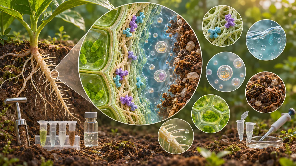
Proteins found outside plant cells can help build cell walls, move signals and interact with microbes. But detecting a protein in an extracellular sample does not prove that it was secreted. A review explains why sampling choices matter and offers guidance for selecting methods and interpreting results.
A plant protein found outside a cell might be part of a cell wall, dissolved in fluid between cells, carried in an extracellular vesicle, or present in liquid around a root or seed. These proteins are involved in plant growth, stress responses and interactions with microbes. But finding a protein in an extracellular sample does not, by itself, reveal how it got there. It may have been secreted, released during normal development or leaked from damaged cells during collection.
Pengxi Wang and Martin Černý survey ways to collect and separate plant extracellular proteins, then reanalyze nine publicly available proteomics datasets using a common prediction pipeline. Their goal is to describe patterns in existing data and help researchers choose methods suited to the biological question they want to ask.
Different samples capture different protein populations
“Extracellular” is not one uniform compartment. Apoplastic washing fluid, collected from spaces within plant tissue, can contain soluble proteins as well as vesicles and material associated with the cell wall. Wall-bound proteins generally require extraction from wall material. Root and seed exudates capture substances in surrounding liquid, while xylem and phloem sap are internal transport fluids collected using particular procedures.
Collection can shape what ends up in a sample. Infiltrating leaves to collect apoplastic fluid may dilute it; excessive pressure or centrifugation may damage cells and introduce intracellular proteins. Cutting a stem to collect sap can also release material from damaged tissue. For extracellular vesicles, a high-speed pellet is better described as “EV-enriched” than as pure: other particles may be collected alongside vesicles.
The authors distinguish the proteins recovered from a particular sample—the extracellular proteome—from the secretome, meaning proteins actively released through secretion pathways. The two are not necessarily the same.
A shared analysis, with important caveats
The authors applied six protein-prediction tools to nine datasets covering leaf apoplastic fluid, root and seed exudates, vascular sap, cell-culture medium, a cell-wall preparation and an EV-enriched fraction. They sorted proteins into four predicted groups: candidates for canonical secretion, candidates for unconventional secretion, intracellular-like proteins, and proteins that did not fit those categories.
In seven datasets, unconventional-secretion candidates outnumbered canonical candidates both by protein count and by relative abundance. This pattern was not universal: suspension-cell culture medium had more canonical than unconventional candidates, while root exudates had similar proportions of the two. Separately, in seven datasets the combined share of canonical and unconventional candidates was higher when measured by relative abundance than when measured by protein count. Seed exudates and the cell-wall preparation showed the opposite pattern. These comparisons describe the analyzed fractions; they do not establish a general rule for all plant extracellular proteins.
The categories are predictions, not direct measurements of secretion routes. A protein without a predicted signal peptide is not thereby proven to use an unconventional pathway. Likewise, an intracellular-like prediction is not a measured contamination rate: proteins can have multiple locations or functions, and some may reach samples through regulated release or association with extracellular structures. A substantial “other” category in one dataset also shows that the classification does not resolve every protein.
Match the method to the question
The review presents a decision tree linking four broad targets—soluble apoplastic proteins, vesicle-associated proteins, wall-associated proteins, and proteins in exudates, sap or culture medium—to possible collection and fractionation strategies. It recommends checks tailored to the sample. These can include monitoring tissue integrity and intracellular marker enzymes for leaf washing fluid, documenting wall enrichment and washing, and using particle measurements, markers and other controls for EV preparations.
Researchers might also compare an extracellular fraction with a whole-tissue sample, collect sequential fluid fractions, or test how protein profiles change as extraction becomes more forceful. Such approaches can offer clues about leakage, but no single check establishes the release route of every protein. Independent localization or functional experiments may be needed for proteins of particular interest.
The comparison has limits: the nine datasets differ in species, tissues, developmental stages, treatments, collection methods and proteomics workflows. Their differences cannot isolate the effect of any one method. The authors recommend clearer reporting of sample handling and fractionation and, where feasible, retaining intermediate fractions so recovery and enrichment can be assessed.
This is a methods-focused review and reanalysis, not a new experiment proving how particular proteins are released. It is a bioRxiv preprint posted November 17, 2026, and the version described had not been certified by peer review. Its findings are best read as a practical framework and patterns to investigate, not a universal map of plant secretion.
- plant proteomics
- extracellular proteins
- plant secretion
- apoplast
- extracellular vesicles
- protein sampling