TRANSMISSION // 2026.11.04.764211v1
The Missing Silicon in a Diatom’s Glass Workshop
Source published
A bioRxiv preprint combines fluorescence imaging, cell fractionation and silicon NMR to investigate how diatoms supply material for their silica walls. It finds evidence for a separable intracellular silicon fraction, but does not establish whether cells use that fraction as a reserve.
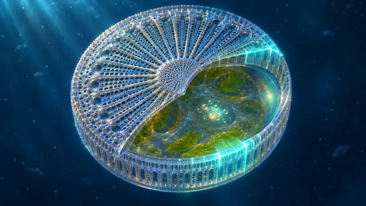
Diatoms can keep building their intricate silica walls even when external silicon becomes scarce. A preprint brings researchers closer to the chemistry of cellular silicon—and exposes a discrepancy that may point to an unidentified source of building material.
Diatoms make glass-like homes from a material they collect from the water. These single-celled algae take up silicic acid and use it to construct patterned silica walls, or frustules. When external silicon runs low, some can continue making new wall material. The question is where they keep the supplies.
A bioRxiv preprint by Tobias Reichelt, Julia Breyer and Eike Brunner approaches that question from two directions: measuring silica made during silicon starvation and trying to separate silicon-containing material from the rest of the cell. The work has not been peer reviewed. Its most intriguing result is not a definitive map of a storage compartment, but a mismatch between what the two approaches appear to find.
A reserve revealed by fluorescence
The researchers studied Cyclotella nana, a marine diatom also widely known by its former name, Thalassiosira pseudonana. They transferred growing cells into artificial seawater without added silicon and supplied PDMPO, a fluorescent marker incorporated into newly forming silica. After 48 hours, more than 99% of cells showed detectable fluorescence, and labeled valve pairs indicated cell division.
That observation supports the existence of a usable silicon reserve: cells were still making silica when the surrounding medium offered little fresh material. Comparing the fluorescence of starved cells with that of fully labeled cells, the authors estimated that the mobilized reserve could represent about 34% of total cellular silicon. Using their measurements of cell silicon content and volume, they translated this to about 540 millimolar averaged over the cell volume—not a direct measurement of dissolved silicic acid.
The number needs caution. It depends on the relationship between fluorescent marker incorporation and silica formation remaining comparable under different silicon conditions. Earlier work cited in the paper suggests that this relationship may change at very low external silicon concentrations. The authors offer an alternative explanation for that earlier finding but treat their own 34% estimate as an upper bound. They also note uncertainty about whether the fully labeled reference captures all stored silicon. Trace silicon was detected in the nominally silicon-free medium, although the authors estimated its contribution per cell was small. Dissolution of existing silica is another potential complication whose rate in living cells was not established.
Four other diatom species yielded fluorescence-based estimates ranging from roughly 4% to 33% of cellular silicon. Each of those species was tested once, so the spread is suggestive rather than a settled comparison across diatoms.
Separating the material
The second approach broke open C. nana cells and sorted their contents through density gradients. The team processed extracted cell walls in parallel as a control: if a silicon-rich fraction appeared in the cell preparation but not in the wall control, it could indicate material originating inside the cells.
Depending on the separation method and sample, the researchers estimated that this cytoplasmic silicon represented about 3–10% of total cellular silicon. They then used silicon-29 enrichment and solid-state nuclear magnetic resonance spectroscopy to examine selected fractions. The spectra were consistent with condensed silica, rather than a large store of simple dissolved silicic acid. The separated material was less highly condensed than the whole-cell sample, which was dominated by wall silica.
This is a chemical clue, not a picture of a storage organelle. The wall control and the differing spectra support the interpretation that the separated fraction is not simply wall debris, but fractionation cannot by itself reveal exactly where that material sat in an intact cell or prove that it serves as a reserve.
The unresolved gap
The fluorescence estimate is much larger than the fraction recovered by separation. Moreover, in a comparison using cells from the same labeled batch, the separated low-density silicon fraction did not fall appreciably after 24 hours of starvation, even though the fluorescence experiment showed silica formation during starvation.
The authors propose that more than one silicon pool may be involved: a relatively stable, separable cytoplasmic fraction and another usable source that was not isolated. They suggest the missing material might be associated with existing cell walls; large aggregates or compartments are alternatives, although particle-migration calculations led them to favor wall association. Reuse of wall silica itself is also raised as a possibility.
Those are hypotheses, not direct observations of a second pool. The starvation experiments used different measurements and time periods, and the reported analytical uncertainties do not capture all variation between cultures or separation procedures. The gap could therefore reflect biology, limitations of the methods, or both.
The preprint makes that gap experimentally tangible. It provides evidence for a separable, chemically condensed silicon fraction that likely originates inside the cells, but its near-constant amount during starvation leaves its role as a usable reserve unproven. The larger question remains open: where does the silicon used to build a new wall come from, and might some of it already be associated with the old one?
- diatoms
- Cyclotella nana
- silicon storage
- biosilica
- fluorescence microscopy
- density gradient centrifugation
- silicon-29 NMR
- preprint