TRANSMISSION // 2026.11.03.759069v1
How a chloroplast transporter helps shape the air inside a leaf
Source published
A bioRxiv preprint links loss of the amino acid transporter RETICULATA to reduced early cell proliferation and enlarged air spaces in Arabidopsis palisade tissue. The findings challenge a cell-death explanation, but the molecular connection between chloroplast transport and leaf development remains unresolved.
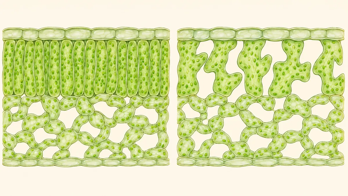
Leaves depend on the spaces between their cells as well as the cells themselves. By following a mutant through leaf development, researchers have traced unusually large air spaces to changes in cell division and shape—not to a detectable increase in dying cells.
A leaf’s internal architecture combines living cells with air spaces that allow carbon dioxide to move through its photosynthetic tissues. But how developing leaves establish that architecture remains poorly understood. A new study examines a chloroplast-associated protein whose loss changes the balance between cells and air, particularly in the tightly packed tissue beneath the upper leaf surface.
In a bioRxiv preprint posted on November 4, 2026, Edwyn A. Locke Beviá and colleagues report that Arabidopsis plants lacking functional RETICULATA, or RE, show reduced early proliferation of palisade mesophyll cells, followed by larger, more irregular cells. Their evidence supports a developmental explanation for the mutant’s enlarged air spaces rather than an explanation based on increased cell death. The work has not been peer reviewed.
Two tissues, different responses
Leaves commonly contain two contrasting photosynthetic tissues. Palisade mesophyll lies beneath the upper epidermis and typically has closely packed, column-shaped cells. Spongy mesophyll lies closer to the lower surface and contains irregularly shaped cells separated by larger gaps.
RE is an amino acid transporter in the inner chloroplast envelope, a function established in earlier research cited by the authors. Plants carrying the loss-of-function mutation re-6 have a distinctive appearance: dark green veins stand out against a pale green leaf blade. This study asked how their unusual internal spaces develop.
The researchers compared the first pair of true leaves in mutant and wild-type plants, using fluorescent staining and confocal microscopy across development. They measured porosity as the proportion of a two-dimensional image region occupied by air space—not as a direct measurement of whole-leaf air volume.
In the palisade, mutant porosity became significantly higher from six days after stratification onward. By day 14, it reached about 45%, compared with about 20% in wild-type plants. Both approached a plateau at roughly the same time, and overall leaf area did not differ significantly.
Spongy mesophyll porosity, by contrast, followed a wild-type-like trajectory. Most measured spongy cell traits were also unchanged at day 23, although mutant cells had a slightly higher aspect ratio. The disruption was therefore concentrated in the palisade rather than equally affecting both tissues.
Testing alternative explanations
One proposed explanation for enlarged spaces in reticulata mutants was that cells die and leave gaps behind. The team tested this using Sytox Orange, a dye that stains non-viable cells, at eight and 14 days after stratification. Laser-damaged palisade cells provided a positive control showing that the assay could detect damaged cells in this tissue.
The researchers found no significant increase in dead palisade cells in re-6 at either time point. These observations argue against increased cell death as the explanation under the conditions examined, although they do not monitor every developmental moment.
Stomatal density offered another possible explanation, because earlier studies have connected these epidermal pores with internal air-space development. Most measurements showed no significant density difference between genotypes, with transient exceptions on each leaf surface. Air-space differences also began before stomata opened, making altered stomatal density an unlikely primary explanation.
Nor did the mutant’s increasingly lobed palisade cells show the tested molecular signs of becoming spongy cells. At day 14, they retained expression of the palisade reporter IQD22 and lacked detectable expression of the spongy reporter CORI3. These two markers support retention of palisade identity despite altered shape.
Fewer divisions before larger cells
To assess proliferation, the team induced fluorescent labels in individual cells and counted their descendants three days later. Between days four and seven, mutant sectors averaged 2.59 cells, compared with 3.94 in wild-type sectors. This clonal assay provides evidence of reduced early palisade proliferation.
Cell enlargement became significantly different later, from day 14 onward. By day 23, mutant palisade cells had about 1.8 times the measured area of wild-type cells and more pronounced lobes and irregular boundaries.
The sequence leaves an important uncertainty: later enlargement could compensate for reduced early division, or RE could influence both processes more directly. The study does not distinguish these possibilities.
The biochemical link also remains open. The authors propose that altered amino acid balance could affect polyamines associated with cell proliferation, but they did not test that pathway here. Their suggestion that RE connects light conditions to palisade development likewise remains a hypothesis. For now, the strongest result is a developmental connection: loss of this chloroplast transporter changes early cell proliferation and later cell morphology alongside a marked increase in palisade air space.
- RETICULATA
- Arabidopsis thaliana
- chloroplast
- palisade mesophyll
- leaf development
- intercellular air spaces
- cell proliferation